Abstract
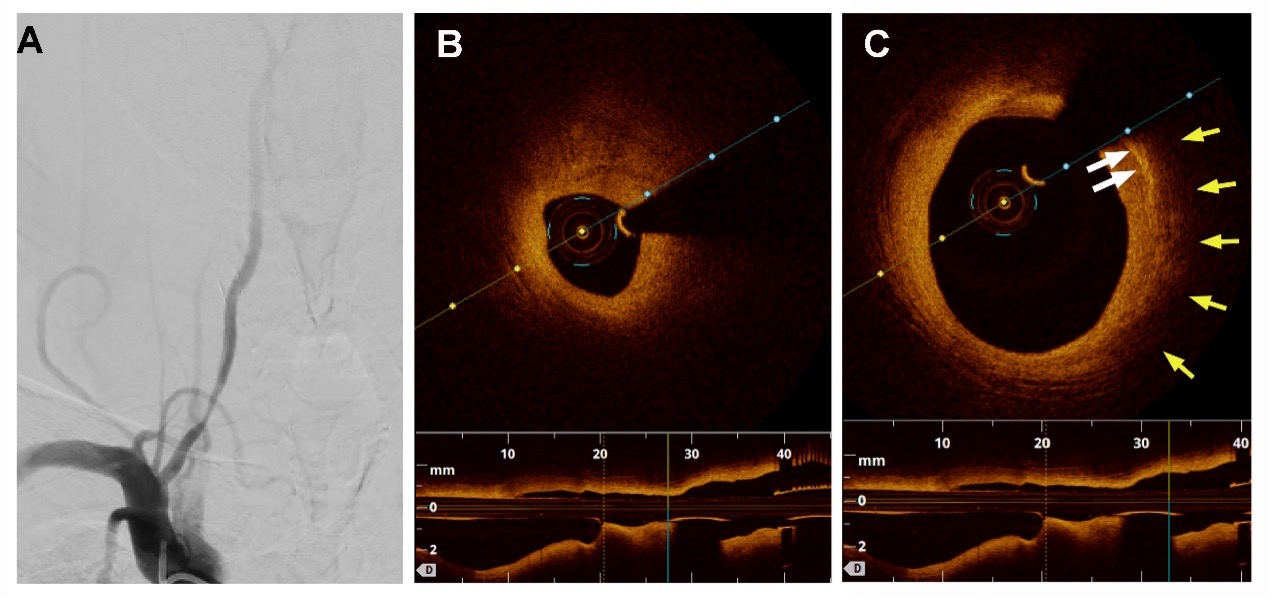
Background: Optical Coherence Tomography (OCT) in a new technology with potential neurointerventional applications. We report the use of OCT to detect intraluminal dissection secondary to angioplasty in the extracranial vertebral artery.
Methods: OCT were acquired using Dragonfly imaging catheter (Dragonfly Duo;LightLab Imaging, Inc.,St.Jude Medical.) and data were analyzed by the ILUMIEN OPTIS Imaging System (Abbott Medical,Westfort,USA) in cervical vertebral artery in an adult patient presented with a 7-day history of dizziness, vertigo and right limb weakness associated with a long stenotic segment of the proximal right vertebral artery segment.
Results: Angiogram showed good patency and no proximal dissection after drug eluting balloon dilation of the stenosis. However, OCT showed two distinct dissections and a 3.5*35 mm RQES drug eluting stent was placed. OCT showed resolution of dissection and new high signals distributed unevenly on the initial surface, presumably drug particles released from the drug eluting balloon.
Conclusions: The first reported case suggests that OCT is feasible for imaging proximal vertebral artery lesion and may be more sensitive than angiogram in detecting post angioplasty vertebral artery dissection.

This work is licensed under a Creative Commons Attribution-NonCommercial-NoDerivatives 4.0 International License.
Copyright (c) 2022 Journal of Vascular and Interventional Neurology

